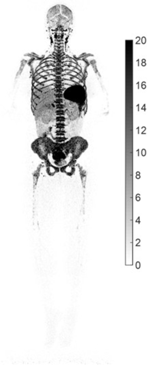
HOUSTON, Aug. 10, 2022 /PRNewswire/ -- UC Davis, a leading university in molecular imaging that collaborated with United Imaging to develop the world's first total-body PET/CT scanner, 'uEXPLORER®,' recently presented their research on COVID-19 at the 2022 Society of Nuclear Medicine and Molecular Imaging (SNMMI) annual meeting. Guided by the uEXPLORER scanner along with a new labeled radiotracer with high affinity to T cells (89Zr-Crefmirlimab-Berdoxam), UC Davis Project Scientist and lead author Dr. Negar Omidvari unveiled the first high-resolution PET image of total-body CD8+ T-cell distribution after COVID-19 infection. The image could shed light on the pathological mechanism and immune response to COVID-19 infection.

The first reported image of CD8+ T cell distribution in a recovered COVID-19 patient (F, 29 y/o, BMI 29), imaged 7 weeks post symptom onset on the uEXPLORER total-body PET/CT scanner, using a ~0.5 mCi injected dose of 89Zr-Df-Crefmirlimab-Berdoxam. Lymphoid organs containing CD8+ T cells (such as spleen, bone marrow, lymph nodes, and tonsils) showed the largest uptake, reflected by the darker black parts of the image. Image courtesy of Dr. Negar Omidvari at UC Davis.
Cytotoxic T cells are key players in the cell-mediated immune response against viral infections. However, 95% of T cells are in tissues rather than in the blood circulation and are therefore difficult to assess non-invasively. High sensitivity PET imaging using a specific T cell radiotracer offers a new methodology to track tissue concentrations of T cells without invasive sampling. The radiotracer used, 89Zr-Crefmirlimab-Berdoxam, is an investigational tracer provided for the study by ImaginAb who is currently in Phase IIb testing to determine predictive performance.
In the reported study, patients recovering from COVID-19 infection received a low injection dose (~0.5 mCi) of 89Zr-labeled radiotracer that targeted CD8+ T cells and underwent a uEXPLORER PET/CT scan at different time points. A control group was also studied. The total-body PET images revealed subtle differences between five recovering COVID-19 patients and three healthy volunteers. The exceptional image quality obtained using the uEXPLORER scanner confirmed CD8+ T cell deposition in the spleen, liver, bone marrow, lymph nodes, and tonsils, as well as differences in the clearance rate of the radiotracer from the blood. Imaging was accomplished with minimal radiation dose to the patients. This research paves the way for further exploration of the assessment of immune response to infectious diseases and cancer, the assessment of response to vaccines in human subjects, and the fundamental mechanisms of the immune system in general.
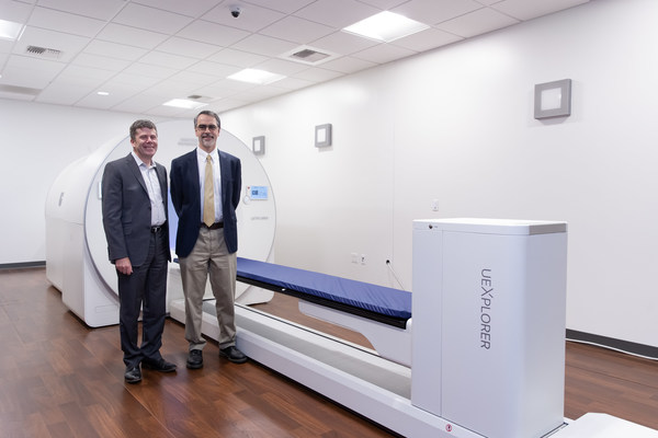
This disruptive technology co-developed by UC Davis and United Imaging was selected as one of the "2018 Breakthroughs of the Year" by Physics World. It pushes the boundaries of traditional PET scanners, with an effective sensitivity that is up to 68-fold greater than current commercial PET scanners. Combined with a 194 cm axial PET field of view, the uEXPLORER scanner can capture dynamic changes to radiotracer distribution across the entire human body with high temporal resolution, which is an essential attribute for this type of research.

Dr. Simon R. Cherry (left) and Dr. Ramsey D. Badawi (right) standing in front of the uEXPLORER scanner.
Simon R. Cherry, Ph.D., Distinguished Professor of Biomedical Engineering and Radiology at UC Davis, was recently awarded the Benedict Cassen Prize at the 2022 SNMMI annual meeting for his pioneering work and immense contributions to nuclear medicine instrumentation and molecular imaging. He commented: "We have now contributed to several different disease areas based on the high sensitivity and total-body coverage of the uEXPLORER scanner. This research is another demonstration of how groundbreaking imaging technology, combined with highly specific radiotracers, can open new opportunities in studying the complex interactions within the whole body, both in health and in response to disease. We hope this research will contribute to a better understanding of the immunological response to COVID-19, and serve as an example for how the immune system can be interrogated at a systems level with total-body PET."
![]() View original content to download multimedia:https://www.prnewswire.com/news-releases/uc-davis-new-research-shows-how-total-body-pet-imaging-can-assess-the-immunological-response-to-covid-19-infections-301603409.html
View original content to download multimedia:https://www.prnewswire.com/news-releases/uc-davis-new-research-shows-how-total-body-pet-imaging-can-assess-the-immunological-response-to-covid-19-infections-301603409.html